Characterizing tau aggregates in neurodegenerative diseases
New research reveals a structural model for amyloid fibrils that could aid in future medicinal interventions for Alzheimer's, CTE, and more.
The microtubule-binding protein tau in neurons of the central nervous system can misfold into filamentous aggregates under certain conditions. These filaments are found in many neurodegenerative diseases such as Alzheimer’s disease, chronic traumatic encephalopathy (CTE), and progressive supranuclear palsy. Understanding the molecular structure and dynamics of tau fibrils is important for designing anti-tau inhibitors to combat these diseases.
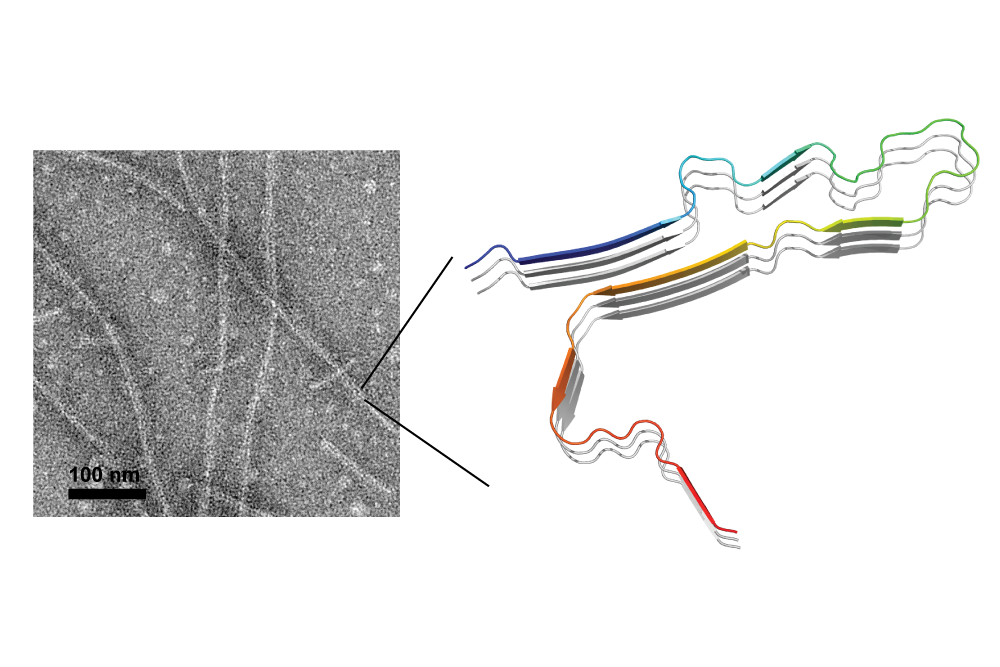
Cryoelectron microscopy studies have recently shown that tau fibrils derived from postmortem brains of Alzheimer’s patients adopt disease-specific molecular conformations. These conformations consist of long sheets, known as beta sheets, that are formed by thousands of protein molecules aligned in parallel. In contrast, recombinant tau fibrillized using the anionic polymer heparin was reported to exhibit polymorphic structures. However, the origin of this in vitro structural polymorphism as compared to the in vivo structural homogeneity is unknown.
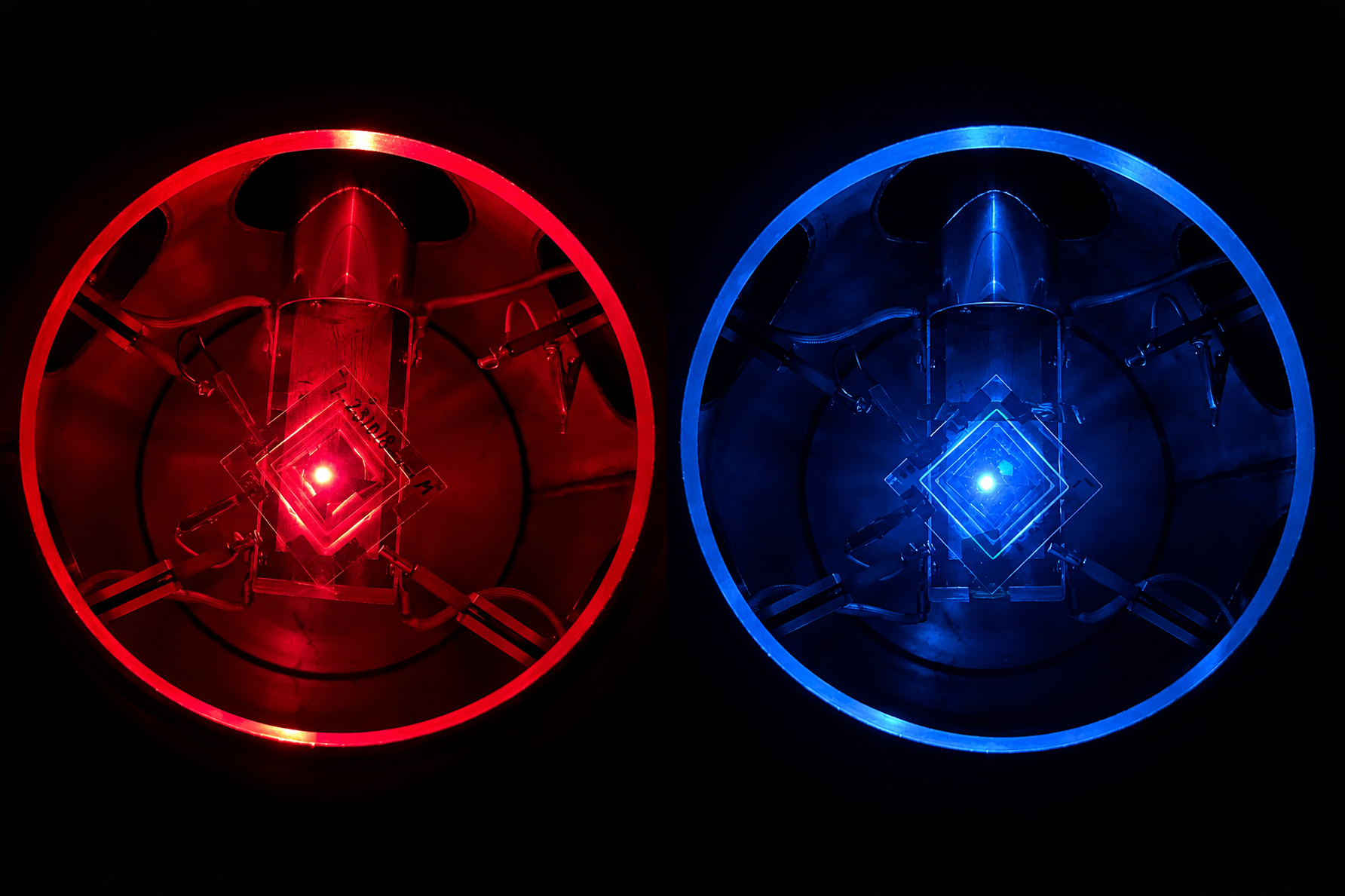
Using solid-state nuclear magnetic resonance (SSNMR) spectroscopy, MIT Professor Mei Hong, in collaboration with Professor Bill DeGrado at the University of California at San Francisco, has shown in a paper, published July 29 in PNAS, that the beta sheet core of heparin-fibrillized tau in fact adopts a single molecular conformation. The tau protein they studied contains four microtubule-binding repeats, and the beta sheet fibril core spans the second and third repeats.
Clarifying biochemical studies of tau and its fibril formation
Previous research on this subject had reported four polymorphic structures of four-repeat (4R) tau fibrils, a polymorphism that led many labs to believe that in vitro tau fibrils were poor mimics of the in vivo patient-brain tau. However, through the use of their SSNMR spectra, which show only a single set of peaks for the protein, Hong and DeGrado discovered a crucial biochemical problem that led to the previous polymorphism.
Once this error was corrected, 4R tau was found to display only a single molecular structure. The revelation of this common biochemical problem, which is protease contamination in the heparin used to fibrillize tau, will significantly clarify and positively impact the field of tau research.
Preventing the formation of tau aggregates in Alzheimer’s disease and beyond
The three-dimensional fold of this four-repeat tau fibril core is distinct from the fibril core of the Alzheimer’s disease tau, which consists of a mixture of three- and four-repeat isoforms. “The tau isoform we studied is the same as that in diseases such as progressive supranuclear palsy, [so] the structural model we determined suggests what the patient brain tau from PSP may look like. Knowing this structure will be important for designing anti-tau inhibitors to either disrupt fibrils or prevent fibrils from forming in the first place,” explains Hong.
This SSNMR study also reported detailed characterizations of the mobilities of amino acid residues outside the rigid beta sheet core. These residues, which appear as a “fuzzy coat” in transmission electron micrographs, exhibit increasingly larger-amplitude motion towards the two ends of the polypeptide chain. Interestingly, the first and fourth microtubule-binding repeats, although excluded from the rigid core, display local b-strand conformations and are semi-rigid.
These structural and dynamical results suggest future medicinal interventions to disrupt or prevent the formation of tau aggregates in some neurodegenerative diseases.