Carly Ziegler wins Koch Institute Image Award
The Koch Institute Image Awards seek to recognize the extraordinary visuals that are produced through life sciences and biomedical research at MIT.
An image contributed by Carly G.K. Ziegler, a graduate student in Professor Alex K. Shalek‘s laboratory, has been selected as one of ten winners of 2019 Koch Institute Image Awards, a competition that seeks to recognize the extraordinary visuals that are produced through life sciences and biomedical research at MIT. Over 160 images were submitted in the competition, and the ten winning images will be exhibited in the large (nearly 8-foot) backlit square and circular displays at the Koch Institute Public Galleries.
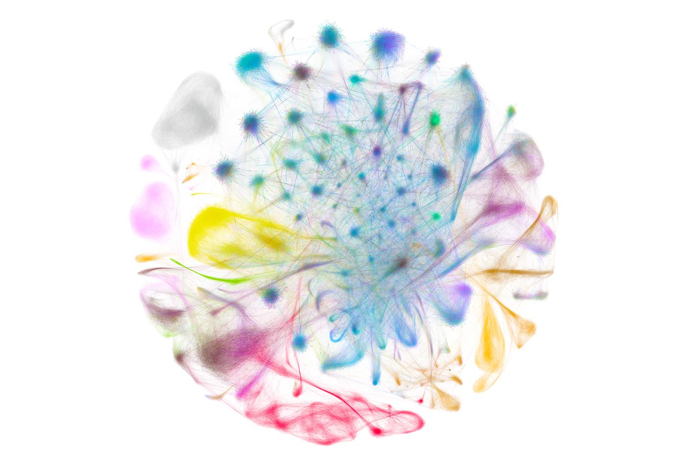
“mRNA is the molecular language cells use to instruct changes to their appearance, function, and interaction with the world,” writes Ziegler in a description of her winning image. “Single-cell RNA-sequencing takes a snapshot of the total mRNA in an individual cell, providing insight into a cell’s current behavior, history, and trajectory. The Shalek lab (MIT – IMES, Chemistry, KI) has pioneered methods for ultra-high-throughput single-cell RNA-sequencing, enabling deep understanding of cellular physiology at an unprecedented scale.”
Ziegler’s winning image represents data comprising the first draft of a Non-Human Primate single cell atlas (45,782 cells, 4 animals, 14 tissues per animal).
“The image is visually striking and, while seemingly simple, provides unprecedented insights into the molecular relationships amongst different cells and compartments.” – Carly G.K. Ziegler
“Single points on the graph represent single cells, their location and the lines between them reflect similarities in their mRNA content,” she explains. “Local areas of high density of points suggest cells of a common phenotype (e.g., activated microglia in the brain), and the point and edge colors reflect the tissue of origin. By characterizing a cell’s “type” purely by its transcriptome, we can validate and redraw cellular taxonomies. Collectively, this analysis allows us to begin to map the full complexity of cellular specialization and diversity in mammals.”
The KI Image Awards are judged by a panel representing a wide range of STEAM disciplines and organizations. Recent judges have included Paula Apsell and Dan Hart, WGBH; Leanne Burden Seidel and Paula Nelson, The Boston Globe; Jonathan Christison and Deborah Sweet, Cell Press; Catherine Draycott, The Wellcome Trust; John Durant, MIT Museum; Carrie Fitzsimmons, ArtScience Labs and Le Laboratorie Cambridge; Andrea Frank, MIT Program in Art, Culture and Technology; Felice Frankel, MIT Center for Materials Science and Engineering; Janis Fraser, Fish & Richardson P.C.; Bethany Millard, Phosphorous Productions; Mary Schneider Enriquez, Harvard Art Museums; Iain Cheeseman and Wendy Salmon, Whitehead Institute for Biomedical Research; previous award-winners and current MIT faculty members.