New AI system uncovers hidden cell subtypes, boosts precision medicine
CellLENS reveals hidden patterns in cell behavior within tissues, offering deeper insights into cell heterogeneity—vital for advancing cancer immunotherapy.
In order to produce effective targeted therapies for cancer, scientists need to isolate the genetic and phenotypic characteristics of cancer cells both within and across different tumors because those differences impact how tumors respond to treatment.
Part of this work requires a deep understanding of the RNA or protein molecules each cancer cell expresses, where it is located in the tumor, and what it looks like under a microscope.
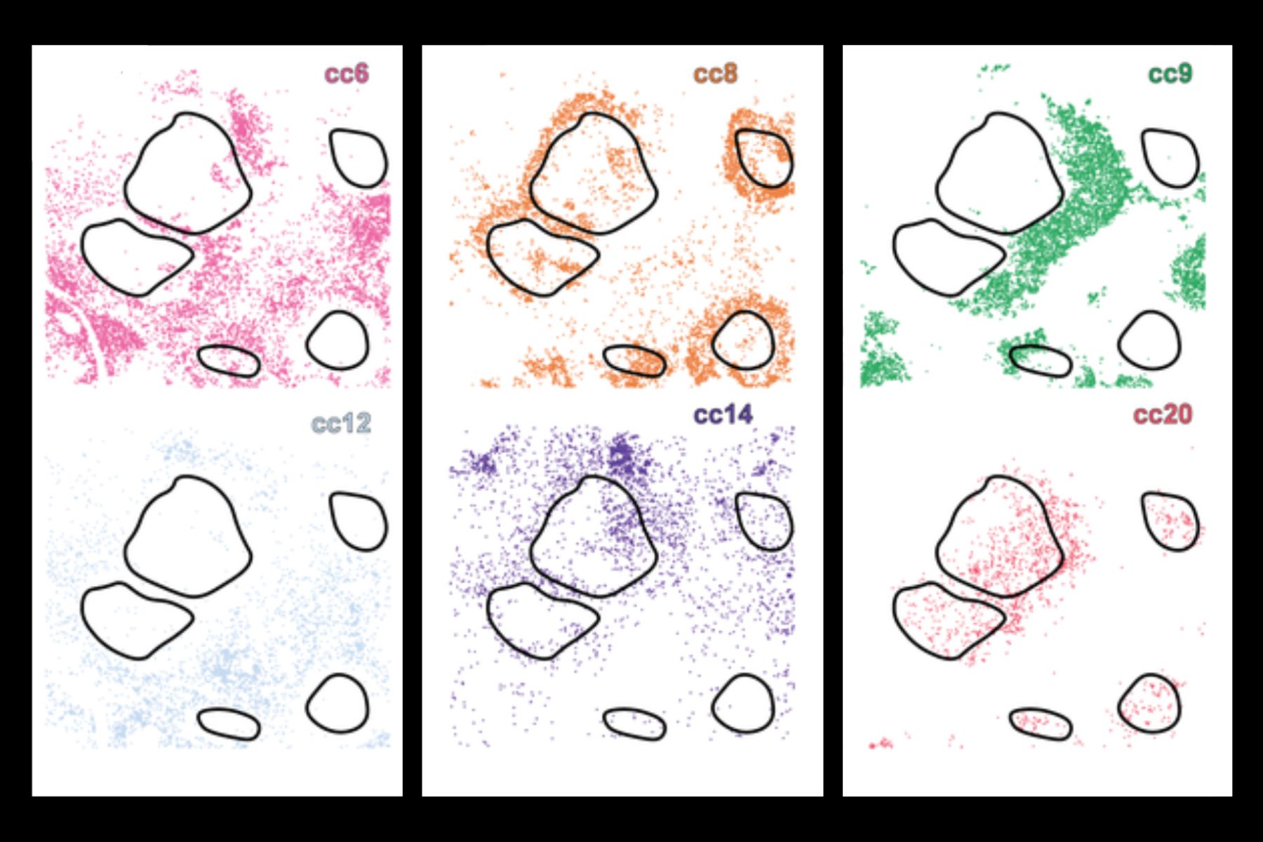
Traditionally, scientists have looked at one or more of these aspects separately, but now a new deep learning AI tool, CellLENS (Cell Local Environment and Neighborhood Scan), fuses all three domains together, using a combination of convolutional neural networks (CNNs) and graph neural networks (GNNs) to build a comprehensive digital profile for every single cell. This allows the system to group cells with similar biology—effectively separating even those that appear very similar in isolation but behave differently depending on their surroundings.
The study, published in Nature Immunology, details the results of a collaboration between researchers from MIT, Harvard Medical School, Yale University, Stanford University, and University of Pennsylvania—an effort led by Bokai Zhu, an MIT postdoc and member of the Broad Institute of MIT and Harvard and the Ragon Institute of MGH, MIT, and Harvard.
Zhu explains the impact of this new tool. “Initially we would say, oh, I found a cell. This is called a T cell. Using the same data set, by applying CellLENS, now I can say this is a T cell and it is currently attacking a specific tumor boundary in a patient.”
“I can use existing information to better define what a cell is, what is the subpopulation of that cell is, what that cell is doing, and what is the potential functional readout of that cell. This method may be used to identify a new biomarker, which provides specific and detailed information about diseased cells, allowing for more targeted therapy development.”
This is a critical advance because current methodologies often miss critical molecular or contextual information—for example, immunotherapies may target cells that only exist at the boundary of a tumor limiting efficacy. By using deep learning, the researchers can detect many different layers of information with CellLENS, including morphology and where the cell is spatially in a tissue.
When applied to samples from healthy tissue and several types of cancer, including lymphoma and liver cancer, CellLENS uncovered rare immune cell subtypes and revealed how their activity and location relate to disease processes—such as tumor infiltration or immune suppression.
These discoveries could help scientists better understand how the immune system interacts with tumors and pave the way for more precise cancer diagnostics and immunotherapies.
“I’m extremely excited by the potential of new AI tools, like CellLENS, to help us more holistically understand aberrant cellular behaviors within tissues. We can now measure a tremendous amount of information about individual cells and their tissue contexts with cutting-edge, multi-omic assays.
Effectively leveraging that data to nominate new therapeutic leads is a critical step in developing improved interventions. When coupled with the right input data and careful downsteam validations, such tools promise to accelerate our ability to positively impact human health and wellness,” says Alex K. Shalek, the Director of the Institute for Medical Engineering & Science (IMES), the J. W. Kieckhefer Professor in IMES & Chemistry, and an extramural member of the Koch Institute at MIT, as well as an Institute Member of the Broad Institute and a Member of the Ragon Institute.